
More Information
Submitted: July 06, 2026 | Approved: July 23, 2026 | Published: July 24, 2026
Citation: Beslin LG. Biochemical Responses to Fenvalerate Exposure in Zebrafish (Danio rerio Hamilton, 1822). Int J Clin Microbiol Biochem Technol. 2026;9(1): 55-60. Available from:
https://dx.doi.org/10.29328/journal.ijcmbt.1001041
DOI: 10.29328/journal.ijcmbt.1001041
Copyright license: © 2026 Beslin LG. This is an open access article distributed under the Creative Commons Attribution License, which permits unrestricted use, distribution, and reproduction in any medium, provided the original work is properly cited.
Keywords: Aquatic toxicity; Mortality; Biochemical alterations; Protein content; Lipid content; SDS–PAGE; Gill; Muscle

Biochemical Responses to Fenvalerate Exposure in Zebrafish (Danio rerio Hamilton, 1822)
Leena Grace Beslin*
Department of Zoology, Nesamony Memorial Christian College (Affiliated to Manonmaniam Sundaranar University), Marthandam-629165, Kanyakumari District, Tamil Nadu, India
*Corresponding author: Leena Grace Beslin, Department of Zoology, Nesamony Memorial Christian College (Affiliated to Manonmaniam Sundaranar University), Marthandam-629165, Kanyakumari District, Tamil Nadu, India. Email: [email protected]
Fenvalerate is a widely used synthetic pyrethroid insecticide with recognised toxicity to aquatic organisms. This study evaluated its toxic effects on zebrafish (Danio rerio) by assessing mortality, biochemical alterations, and protein profile changes following exposure to 20–100 µg/L fenvalerate. Mortality increased in a concentration- and time-dependent manner. At 20 µg/L, mortality increased from 55% at 24 h to 82% at 96 h, while 40 and 60 µg/L resulted in mortality rising from 62% to 94% and 71% to 99%, respectively. Exposure to 80 µg/L caused 79–100% mortality, and complete mortality was observed at 80 and 100 µg/L after 72 and 96 h, respectively. Sublethal (LC50) exposure induced significant biochemical changes in gill and muscle tissues. Gill protein content decreased from 120.0 ± 1.22 mg/g in controls to 110.0 ± 1.40 mg/g after 96 h, whereas muscle protein declined from 80.2 ± 1.3 to 60.0 ± 2.7 mg/g. Lipid reserves were also markedly reduced, with muscle lipid content decreasing from 4.0 ± 0.46% to 3.1 ± 0.12% and gill lipid content from 6.5 ± 0.22% to 2.5 ± 0.31% after 96 h. SDS-PAGE analysis revealed distinct alterations in protein banding patterns between control and treated fish, indicating fenvalerate-induced changes in protein expression. These findings demonstrate that fenvalerate causes severe concentration- and time-dependent toxicity, leading to increased mortality, depletion of proteins and lipids, and altered protein profiles, highlighting its ecological risk to aquatic organisms and the utility of zebrafish as a sensitive model for pesticide toxicity assessment.
The widespread use of pesticides has become an indispensable component of modern agricultural practices to improve crop productivity and protect against pest infestations. Despite their agricultural benefits, pesticides frequently enter aquatic ecosystems through agricultural runoff, leaching, and drainage, posing significant risks to non-target organisms, particularly fish [1]. Environmental factors such as temperature, pH, and dissolved oxygen can further influence pesticide persistence and toxicity, thereby increasing their harmful effects on aquatic ecosystems. In addition to pesticides, pollutants such as heavy metals and organic contaminants contribute to the deterioration of water quality and threaten aquatic biodiversity [2].
Fish are widely recognised as valuable bioindicators for monitoring environmental pollution because they respond sensitively to changes in water quality and readily accumulate toxic substances from their surroundings [3]. Exposure occurs either directly through contaminated water or indirectly through the aquatic food chain [4]. Consequently, physiological and biochemical alterations in fish provide important information regarding the ecological impact of environmental contaminants.
Among the different classes of pesticides, insecticides have received considerable attention because of their adverse effects on aquatic flora and fauna. A substantial proportion of applied insecticides fails to reach the intended target organisms and is transported into nearby rivers, lakes, and ponds through surface runoff. Continuous discharge of agricultural and industrial pollutants into freshwater ecosystems has become a major environmental concern worldwide [5]. The extensive and often indiscriminate use of pesticides has therefore prompted researchers to investigate their ecological consequences and their impact on aquatic organisms [6].
Synthetic pyrethroids are among the most extensively used insecticides because of their high insecticidal efficacy, low persistence in terrestrial environments, and relatively low mammalian toxicity. However, these compounds are highly toxic to aquatic organisms, particularly fish, owing to their limited ability to metabolise and eliminate pyrethroids [7]. Contamination of aquatic habitats with pyrethroids not only threatens fish populations but may also have indirect consequences for human health through the aquatic food web, as fish constitute an important source of dietary protein.
Several studies have demonstrated the toxic effects of pyrethroid pesticides in zebrafish (Danio rerio). Tarkhani, et al. [8] reported that exposure to diazinon and deltamethrin caused concentration- and time-dependent mortality in zebrafish, with LC₅₀ values confirming their acute toxicity. Similarly, Sapanadevi and Gupta, [9] observed that the sensitivity of zebrafish to lindane, deltamethrin, and atrazine varied with developmental stage, with larvae exhibiting greater susceptibility to deltamethrin and atrazine than fingerlings. Mohapatra, et al. [10] further demonstrated that fenvalerate exposure significantly reduced the total protein content in Labeo rohita, whereas dietary probiotic supplementation alleviated the toxic effects.
Fenvalerate is a broad-spectrum synthetic pyrethroid extensively used in agriculture, animal husbandry, and public health programs. Although highly effective against insect pests, fenvalerate frequently reaches freshwater environments through agricultural runoff, where it poses a serious threat to aquatic organisms. Fish are particularly vulnerable because of their relatively low capacity to detoxify and excrete pyrethroid compounds, making them highly sensitive even to low concentrations of fenvalerate [11]. Toxic exposure can impair growth, metabolism, immune function, and other physiological processes, ultimately affecting fish survival and ecosystem health.
In view of these concerns, the present study was undertaken to evaluate the toxic effects of fenvalerate on zebrafish (Danio rerio). The study investigated concentration- and time-dependent mortality following acute exposure and assessed the biochemical responses by measuring protein and lipid contents in the gill and muscle tissues. In addition, alterations in protein expression were examined using SDS–PAGE analysis. The findings of this study provide valuable insights into the biochemical and molecular responses of zebrafish to fenvalerate exposure and contribute to a better understanding of the ecological risks associated with pyrethroid contamination in freshwater ecosystems.
Experimental animals
Healthy adult zebrafish (Danio rerio) were procured from a commercial aquarium facility in Nagercoil, Kanyakumari District, Tamil Nadu, India. The fish were transported to the laboratory in aerated containers to minimise transportation stress and were allowed to acclimatise before the commencement of the experiments.
Acclimatisation and maintenance
The experimental fish were acclimatised under laboratory conditions for 15 days before exposure. Fish were maintained in glass aquaria (60 × 30 × 25 cm) containing dechlorinated freshwater under a 12 h light/12 h dark photoperiod. Water temperature was maintained at 28 ± 2°C, and continuous aeration was provided throughout the experimental period. Approximately 20 fish were maintained in each aquarium. Water was renewed daily to maintain optimum water quality, and fish were fed twice daily with freeze-dried Tubifex worms. Feeding was discontinued 24 h before the toxicity experiment. All experiments were performed in triplicate.
Acute toxicity assessment of fenvalerate
Technical-grade fenvalerate (99.9% purity; Merck, Bangalore, India) was used as the test pesticide. After acclimatisation, fish were randomly divided into one control group and five treatment groups. The treatment groups were exposed to fenvalerate concentrations ranging from 20 to 100 µg/L, while the control group was maintained under identical conditions without pesticide exposure. Acute toxicity tests were conducted for 96 h, and mortality was recorded after 24, 48, 72, and 96 h of exposure. Dead fish were removed immediately after observation, and cumulative mortality (%) was calculated according to standard acute toxicity testing procedures [12].
Sublethal exposure experiment
For biochemical analyses, zebrafish with an average body length of 3.0 ± 0.21 cm and body weight of 0.31 ± 0.02 g were selected. Fish were acclimatised for two weeks and maintained under identical laboratory conditions. The experimental group was exposed to a sublethal concentration equivalent to one-tenth of the 96-h LC₅₀ value of fenvalerate for one week, whereas the control group remained in pesticide-free water. Following exposure, fish were sacrificed, and gill and muscle tissues were excised for biochemical and protein expression analyses.
Estimation of total protein
The total protein content of the gill and muscle tissues was determined using the Lowry method [13]. Briefly, tissue samples were homogenised in an appropriate buffer, and proteins were reacted with alkaline copper reagent followed by the Folin–Ciocalteu phenol reagent to produce a blue-colored complex. The absorbance was measured spectrophotometrically, and protein concentration was expressed as mg/g tissue.
Estimation of total lipid
Total lipid content was estimated using the chloroform–methanol extraction method (2:1, v/v) described by Mahmoud, et al. [14], which is based on the classical extraction procedure of Folch, et al. [15]. Approximately 0.1 g of gill and muscle tissues was washed thoroughly with distilled water, dried at 60°C for 24 h, homogenised with chloroform: methanol (2:1, v/v), and filtered. Phase separation was achieved by adding saline solution, after which the organic phase containing lipids was collected. The solvent was evaporated, and the extracted lipid was weighed. Lipid content was expressed as the percentage of wet tissue weight.
SDS–PAGE analysis
Protein expression profiles in the gill and muscle tissues were analysed by sodium dodecyl sulfate–polyacrylamide gel electrophoresis (SDS–PAGE) according to the method of Laemmli, [16]. Protein samples were denatured with SDS to impart a uniform negative charge and separated according to molecular weight using polyacrylamide gel electrophoresis. Equal quantities of protein from control and fenvalerate-treated fish were loaded onto the gel. After electrophoresis, protein bands were visualised and compared to evaluate changes in protein expression induced by fenvalerate exposure.
Statistical analysis
All experiments were conducted in triplicate, and the results are presented as mean ± standard deviation (SD). Significant differences between the control and treatment groups were determined by error bars, and P < 0.05 was considered statistically significant [17].
Acute toxicity of fenvalerate in zebrafish
Acute exposure to fenvalerate caused a marked increase in mortality of Danio rerio in both a concentration- and time-dependent manner (Figure 1). Fish exposed to 20 µg/L exhibited 55% mortality after 24 h, which increased significantly to 82% after 96 h (p < 0.05). Likewise, mortality at 40 µg/L increased from 62% at 24 h to 94% after 96 h. At 60 µg/L, mortality increased from 71% during the initial 24 h to 99% by the end of the exposure period. Exposure to 80 µg/L resulted in mortality ranging from 79% to 100%, while complete mortality (100%) was observed at both 80 and 100 µg/L after 72 and 96 h, respectively. Statistical analysis using standard deviation and error bars revealed significant differences in mortality among the exposure concentrations and durations (p < 0.05), indicating that mortality increased significantly with increasing fenvalerate concentration and exposure time.
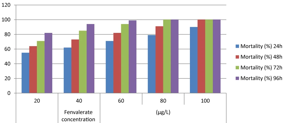
Figure 1: Effect of different (20 – 100 µg/L) concentrations of fenvalerate on Zebrafish at various exposure times.
Effect of fenvalerate on total protein content
Gill protein: Fenvalerate exposure caused a progressive reduction in the total protein content of zebrafish gills (Figure 2). The control fish exhibited a protein content of 120.0 ± 1.22 mg/g, whereas exposed fish showed values of 118.31 ± 0.50, 117.40 ± 1.80, 114.30 ± 2.10, and 110.00 ± 1.40 mg/g after 24, 48, 72, and 96 h, respectively.
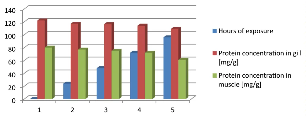
Figure 2: Total protein content of muscle and gill of Zebrafish.
Muscle protein: A similar trend was observed in muscle tissue. The protein concentration decreased from 80.2 ± 1.3 mg/g in the control fish to 78.4 ± 2.4, 71.4 ± 2.2, and 60.0 ± 2.7 mg/g following fenvalerate exposure, with the lowest value recorded after 96 h. Statistical analysis confirmed a significant reduction in muscle protein content in exposed fish compared with the control group (p < 0.05). Overall, the reduction in protein concentration was more pronounced in muscle tissue after prolonged exposure, suggesting disruption of protein metabolism under fenvalerate-induced stress.
Effect of fenvalerate on total lipid content
Fenvalerate exposure significantly altered the lipid reserves of zebrafish (Figure 3). The muscle lipid content decreased from 4.0 ± 0.46% (wet weight) in the control group to 3.1 ± 0.12% after 96 h of exposure (p < 0.05). A greater reduction was observed in gill tissue, where lipid content declined from 6.5 ± 0.22% in the control group to 2.5 ± 0.31% after 96 h. Statistical comparison revealed that both muscle and gill lipid contents were significantly reduced following fenvalerate exposure compared with the control (p < 0.05). The greater depletion of lipids in gill tissue indicates that the gill is more susceptible to fenvalerate-induced biochemical stress.
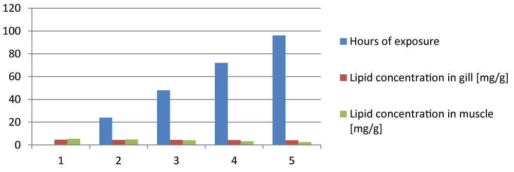
Figure 3: Lipid content of muscle and gill of Zebrafish.
SDS–PAGE analysis of protein expression
SDS–PAGE analysis demonstrated distinct alterations in the protein banding patterns of fenvalerate-exposed zebrafish compared with the control group (Figure 4). Protein bands in the exposed gill and muscle tissues exhibited reduced staining intensity, while several bands appeared to be absent or newly expressed following pesticide exposure. These changes suggest that fenvalerate interfered with normal protein synthesis and induced stress-associated protein expression. The observed modifications in protein profiles indicate alterations in gene expression and protein metabolism resulting from induced pesticide physiological stress.
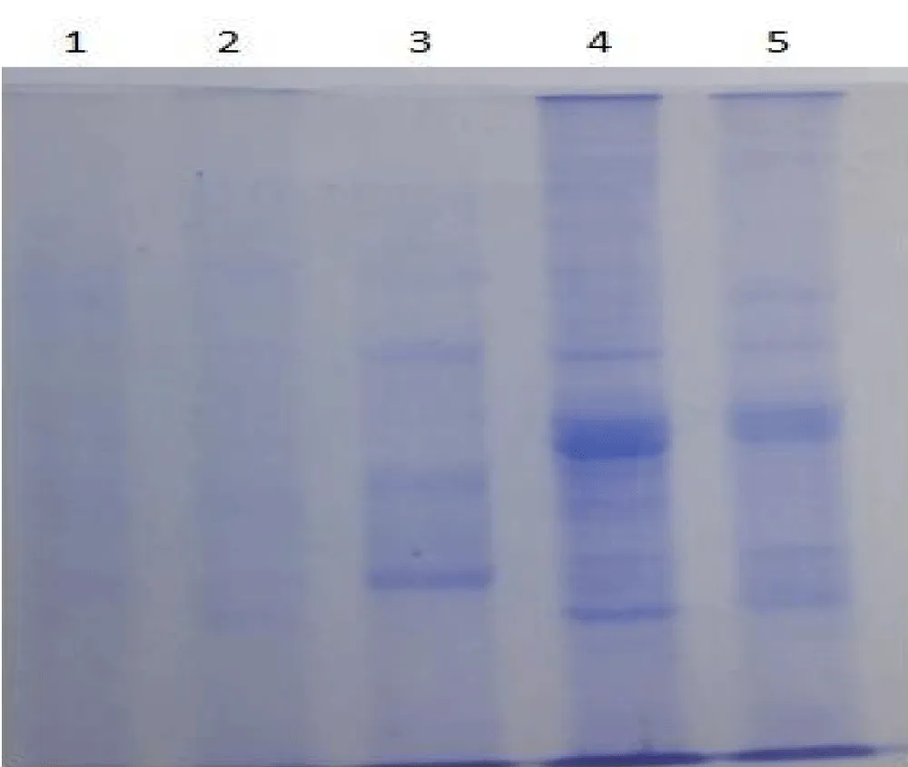
Figure 4: SDS-PAGE: Changes in protein bands in gill and muscle of zebrafish. 1- Normal gill, 2 –Fenvalerate-exposed gill, 3 – Protein marker, 4 – Normal muscle, 5 – Fenvalerate-exposed muscle.
Statistical analysis
Data are presented as mean ± standard deviation (SD) of three independent experiments with error bars. Differences between the control and fenvalerate-treated groups were considered statistically significant at p < 0.05.
The present study demonstrates that fenvalerate exerts pronounced toxic effects on Danio rerio, as evidenced by concentration- and time-dependent mortality, depletion of tissue proteins and lipids, and alterations in protein expression profiles. These findings indicate that fenvalerate disrupts normal physiological and metabolic functions, thereby compromising the health and survival of exposed fish. Acute toxicity assays showed that mortality increased progressively with increasing fenvalerate concentration and exposure duration. Complete mortality was observed at the highest concentrations after prolonged exposure, confirming the high sensitivity of zebrafish to this pyrethroid insecticide.
Synthetic pyrethroids are known to interfere with voltage-gated sodium channels in nerve cells, resulting in prolonged neuronal excitation, impaired swimming behaviour, respiratory distress, and eventually death [18]. Because fish possess a limited capacity to metabolise pyrethroids, even low environmental concentrations can produce severe toxic effects. The concentration-dependent mortality observed in the present study agrees with previous investigations that reported acute toxicity of fenvalerate and related pyrethroids in freshwater fishes, including Cyprinus carpio, Labeo rohita, and Danio rerio [7,19].
Proteins constitute essential structural and functional components of living cells and are involved in enzymatic reactions, tissue growth, immune defence, and cellular repair. In the present investigation, a progressive reduction in protein content was observed in both gill and muscle tissues following fenvalerate exposure. Protein depletion under pesticide stress is generally attributed to enhanced proteolysis and the utilisation of amino acids as alternative energy sources when normal carbohydrate metabolism is disturbed. Toxic stress may also inhibit protein synthesis by affecting ribosomal activity and gene expression, resulting in reduced tissue protein levels. Similar reductions in protein concentration have been reported in fish exposed to pyrethroids and other pesticides, suggesting that protein depletion is a reliable biochemical indicator of toxic stress [20,21]. The greater decline observed after prolonged exposure indicates that the metabolic burden increases with exposure duration.
Lipid reserves also decreased markedly in both gill and muscle tissues after fenvalerate exposure. Lipids represent an important source of metabolic energy and contribute to membrane integrity and cellular signalling. Under toxic conditions, stored lipids are mobilised to satisfy the increased energy demand associated with detoxification and stress adaptation. Furthermore, pesticide-induced oxidative stress can initiate lipid peroxidation, resulting in degradation of membrane lipids and disruption of cellular structure. The pronounced depletion of lipids in the gills observed in this study may reflect the direct contact of this organ with contaminated water, making it particularly susceptible to pesticide uptake and oxidative injury. Comparable reductions in tissue lipid content have been documented in pesticide-exposed freshwater fishes, including Cyprinus carpio, Labeo rohita, and Clarias gariepinus [22,23]. Recent studies have also shown that pyrethroid exposure disrupts lipid metabolism through oxidative damage and mitochondrial dysfunction [24].
The gills are among the first organs affected by waterborne toxicants because of their extensive surface area and direct contact with the aquatic environment. During the exposure period, behavioural abnormalities such as irregular swimming, excessive mucus secretion, and increased opercular movement were observed. These responses are characteristic indicators of respiratory stress and reduced physiological performance in pesticide-exposed fish. Excessive mucus production may represent a protective mechanism to reduce toxicant entry; however, it can simultaneously impair gas exchange and increase respiratory effort. Similar behavioural responses have been reported in fish exposed to pyrethroids and organophosphate pesticides, supporting the use of behavioural endpoints as early indicators of aquatic pollution [25,26].
SDS–PAGE analysis revealed noticeable changes in protein banding patterns between control and fenvalerate-exposed fish. Reduced intensity of several protein bands together with the appearance or disappearance of specific bands suggests that fenvalerate alters protein expression. These changes may result from inhibition of normal protein synthesis, degradation of existing proteins, or induction of stress-responsive proteins that help cells adapt to toxic conditions. Alterations in protein expression have been widely recognised as molecular biomarkers of environmental stress because they reflect changes in gene regulation and cellular metabolism. Similar observations have been reported in fish exposed to pyrethroids and other environmental contaminants, where electrophoretic analyses demonstrated modifications in proteins associated with antioxidant defence, metabolism, and cellular protection [27]. Recent proteomic investigations in zebrafish have further confirmed that pesticide exposure alters pathways related to energy metabolism, protein folding, oxidative stress, apoptosis, and immune regulation [28,29].
The biochemical alterations observed in this study indicate that fenvalerate exposure disrupts metabolic homeostasis in zebrafish. Depletion of proteins and lipids suggests increased energy expenditure to maintain physiological functions under chemical stress, while alterations in protein expression demonstrate activation of cellular defence mechanisms. Together, these responses highlight the sensitivity of zebrafish as a model organism for evaluating pesticide toxicity and support the use of biochemical biomarkers in aquatic environmental monitoring.
The findings demonstrate that fenvalerate poses a significant hazard to freshwater fish by inducing concentration- and time-dependent mortality, disrupting energy metabolism, and altering protein expression. Given the widespread use of pyrethroid insecticides in agriculture, continuous monitoring of pesticide residues in aquatic ecosystems is essential to minimise ecological risks. Future studies integrating oxidative stress biomarkers, antioxidant enzyme activities, histopathological examination, and transcriptomic or proteomic analyses would provide a more comprehensive understanding of the mechanisms underlying fenvalerate toxicity.
Limitations
The present study provides valuable information on the acute toxic effects of fenvalerate in zebrafish; however, several limitations should be acknowledged. First, the investigation was conducted under controlled laboratory conditions, which may not fully represent the complex environmental conditions encountered in natural aquatic ecosystems. Second, the study focused primarily on acute exposure and short-term biochemical responses, whereas chronic exposure at environmentally relevant concentrations may produce different physiological and ecological effects. Third, only total protein, total lipid, and SDS-PAGE protein profiling were evaluated. Additional biomarkers, including antioxidant enzymes, oxidative stress indices, neurotransmitter activity, histopathological alterations, and reproductive parameters, would provide a more comprehensive understanding of fenvalerate toxicity. Furthermore, molecular approaches such as gene expression analysis, proteomics, and metabolomics were not included and could better elucidate the mechanisms underlying pesticide-induced toxicity. Finally, although zebrafish are an established model organism for aquatic toxicology, caution should be exercised when extrapolating these findings to other fish species and natural populations with different ecological and physiological characteristics.
The present investigation demonstrates that fenvalerate exerts pronounced toxic effects on zebrafish (Danio rerio) in a concentration- and time-dependent manner. Acute exposure resulted in progressive mortality, while sublethal exposure caused significant depletion of protein and lipid reserves in gill and muscle tissues, indicating disruption of normal metabolic processes. SDS-PAGE analysis further revealed alterations in protein expression, suggesting that fenvalerate induces cellular stress and interferes with protein metabolism. Collectively, these findings establish proteins, lipids, and protein expression profiles as sensitive biomarkers for assessing fenvalerate-induced toxicity in aquatic organisms. The study also confirms the suitability of zebrafish as a reliable model for evaluating pesticide toxicity and environmental risk. Considering the extensive agricultural use of fenvalerate, continuous monitoring of pesticide contamination in freshwater ecosystems and the implementation of sustainable pesticide management strategies are essential to minimise ecological damage and protect aquatic biodiversity. Future investigations integrating biochemical, histopathological, molecular, and omics-based approaches under chronic exposure conditions will provide a more comprehensive understanding of the mechanisms of fenvalerate toxicity and support improved environmental risk assessment.
- Todd NE, Van Leeuwen M. Effects of carbaryl (Sevin) on early life stages of zebrafish. Ecotoxicol Environ Saf. 2000;53:267‑72. Available from: https://doi.org/10.1006/eesa.2002.2231
- Graham R, Scott KA, Sloman SL. The effect of environmental pollutants on complex fish behaviour: integrating behavioural and physiological indicators of toxicity. Aquat Toxicol. 2004;68:369‑92. Available from: https://doi.org/10.1016/j.aquatox.2004.03.016
- Dautremepuits C, Paris‑Palacios S, Betoulle S, Vernet G. Modulation of hepatic and head kidney parameters of carp (Cyprinus carpio) induced by copper and chitosan. Comp Biochem Physiol C Toxicol Pharmacol. 2004;137:325‑33. Available from: https://doi.org/10.1016/j.cca.2004.03.005
- Ateeq B, Abul Farah M, Niamat Ali M, Ahmad W. Induction of micronuclei and erythrocyte alterations in the catfish Clarias batrachus by 2,4‑dichlorophenoxyacetic acid and butachlor. Mutat Res. 2002;518:135‑44. Available from: https://doi.org/10.1016/s1383-5718(02)00075-x
- Gupta PK. Pesticide exposure: Indian scenario. J Appl Toxicol. 2004;24:83‑98. Available from: https://doi.org/10.1016/j.tox.2004.01.021
- Relyea RA. The impact of insecticides and herbicides on the biodiversity and productivity of aquatic communities. Ecol Appl. 2005;15(2):618‑27.
- Richterová Z, Svobodová Z. Pyrethroids and their influence on fish. Slov Vet Res. 2012;49:63‑72.
- Tarkhani R, Hedayati A, Bagheri T, Shadi A, Khalili M. Determination of LC₅₀, NOEC and LOEC of pesticides in zebrafish (Danio rerio). Iran J Comp Clin Pathol Res. 2012;7:57‑62.
- Sapanadevi M, Gupta A. Sublethal toxicity of pyrethroids on biochemical constituents in Anabas testudineus. Pestic Biochem Physiol. 2014;115:48‑52. Available from: https://doi.org/10.1016/j.pestbp.2014.08.004
- Mohapatra S, Chakraborty T, Prusty AK, Das P, Paniprasad K, Mohanta KN. Use of microbial probiotics in the diet of Labeo rohita fingerlings. Aquac Nutr. 2012;18:1‑11.
- Madan VK, Singh R, Kumari B, Naresh JS, Kathpal TS. Dissipation of lindane and fenvalerate residues in chickpea (Cicer arietinum L.) under Indian climatic conditions. Ecotoxicol Environ Saf. 2000;46:163‑6. Available from: https://doi.org/10.1006/eesa.1999.1903
- Organisation for Economic Co‑operation and Development. Test No. 203: Fish, acute toxicity test. OECD guidelines for the testing of chemicals. Paris: OECD Publishing; 2019.
- Lowry OH, Rosebrough NJ, Farr AL, Randall RJ. Protein measurement with the Folin phenol reagent. J Biol Chem. 1951;193:265‑75. Available from: https://pubmed.ncbi.nlm.nih.gov/14907713/
- Mahmoud AH, Darwish NM, Kim YO, Vijayaraghavan P, Kwon JT, Na SW, Kim HJ. Fenvalerate‑induced toxicity in zebrafish (Danio rerio) and biochemical responses. J King Saud Univ Sci. 2020;32(2):1569‑80.
- Folch J, Lees M, Stanley GHS. A simple method for the isolation and purification of total lipids from animal tissues. J Biol Chem. 1957;226:497‑509. Available from: https://pubmed.ncbi.nlm.nih.gov/13428781/
- Laemmli UK. Cleavage of structural proteins during the assembly of the head of bacteriophage T4. Nature. 1970;227:680‑5. Available from: https://doi.org/10.1038/227680a0
- Zar JH. Biostatistical analysis. 5th ed. Upper Saddle River (NJ): Pearson Education; 2010.
- Soderlund DM. Molecular mechanisms of pyrethroid insecticide neurotoxicity.Arch Toxicol . 2012 Feb;86(2):165-81. Available from: https://doi.org/10.1007/s00204-011-0726-x
- Prusty AK, Kohli MPS, Sahu NP, Pal AK, Saharan N, Mohapatra S, Gupta SK. Effect of short‑term exposure to fenvalerate on biochemical and haematological responses in Labeo rohita. Pestic Biochem Physiol. 2011;100:124‑9.
- Mushigeri SB, David M. Accumulation of fenvalerate and changes in enzyme activities in Cirrhinus mrigala. J Basic Clin Physiol Pharmacol. 2004;15:143‑52. Available from: https://doi.org/10.1515/jbcpp.2004.15.3-4.143
- Kumar S, Gopal K. Impact of distillery effluent on physiological consequences in Channa punctatus. Bull Environ Contam Toxicol. 2001;66:617‑22. Available from: https://doi.org/10.1007/s001280053
- Vinodhini R, Narayanan M. Effect of heavy metals on vitamin E, lipid and glycogen reserves in Cyprinus carpio. Maejo Int J Sci Technol. 2008;2:391‑9.
- Hanan SG, El‑Kasheif MA, Ibrahim SA, Authman MMN. Effect of water pollution in El‑Rahawy Drainage Canal on haematology and organs of freshwater fish Clarias gariepinus. World Appl Sci J. 2013;21:329‑41.
- Li X. Oxidative stress and biochemical responses induced by pyrethroid pesticides in fish. Ecotoxicol Environ Saf. 2022;238:113612.
- Pandey S, Parvez S, Sayeed I, Haque R, Bin‑Hafeez B, Raisuddin S. Biomarkers of oxidative stress in Wallago attu. Sci Total Environ. 2003;309:105‑15. Available from: https://doi.org/10.1016/s0048-9697(03)00006-8
- Zutshi BSG, Prasad R, Nagaraja R. Alteration in haematology of Labeo rohita under pollution stress. Environ Monit Assess. 2010;168:11‑9. Available from: https://doi.org/10.1007/s10661-009-1087-2
- Suneetha K, Kumar KG, Veeraiah K. Changes in protein subunits induced by endosulfan and fenvalerate in Labeo rohita through SDS‑PAGE. J Environ Biol. 2010;31(5):759‑63.
- Chen H. Proteomic responses of zebrafish following pesticide exposure. Environ Pollut. 2023;317:120738.
- Zhang L. Molecular mechanisms of pyrethroid toxicity in aquatic organisms. Chemosphere. 2023;327:138516.