More Information
Submitted: 02 March 2020 | Approved: 27 March 2020 | Published: 30 March 2020
How to cite this article: Sharma A, Ponmariappan S. Development of ELISA based detection system against C. botulinum type B. Int J Clin Microbiol Biochem Technol. 2020; 3: 017-020.
DOI: 10.29328/journal.ijcmbt.1001010
Copyright License: © 2020 Sharma A, et al. This is an open access article distributed under the Creative Commons Attribution License, which permits unrestricted use, distribution, and reproduction in any medium, provided the original work is properly cited.
Keywords: Anaerobic; Botulism; ELISA; Centrifugation; Antiserum
Development of ELISA based detection system against C. botulinum type B
Arti Sharma1,2* and S Ponmariappan2
1Government Degree College Prithvipur, Niwari (MP) 472336, India
2Research Scholar, Biotechnology Division, Defence Research & Development Establishment, Gwalior-474 002, India
*Address for Correspondence: Research Scholar, Biotechnology Division, Defence Research & Development Establishment, Gwalior-474 002, India
Botulism is the disease caused by botulinum neurotoxins. It is produced by an obligate anaerobic bacteria called Clostridium botulinum. There is no immuno-detection system available in the world for the detection of C. botulinum. Secretory proteins of cooked meat media grown C. botulinum type B were extracted by TCA precipitation method. Polyclonal antibodies were generated against secretory proteins. Cytokine profiling of secretory proteins were done. An immunodetection system was developed to detect the C. botulinum type B using Secretory proteins of C. botulinum type B.
Botulism is the most dangerous disease caused by botulinum neurotoxin (BoNTs). It is secreted by obligate anaerobic bacteria called Clostridium botulinum. There are eight serotypes of BoNT designated A-H. Out of seven serotypes, A, B, E and F cause the human botulism and remaining serotypes cause the botulism in animals and birds. Mainly there are three types of botulism. The classical form of botulism is food borne botulism, an intoxication that follows the consumption of food containing preformed neurotoxin. Unlike food-borne botulism, the other forms of human botulism (infant and wound botulism) are really infections where the toxigenesis occurs in vivo. In the case of infant and wound botulism, primary infection followed by secondary intoxication. In national scenario (India), botulism outbreak were found in Gujarat , New Delhi and Coimbatore [1,2]. According to CDC report, every year 145 cases of human botulism are reported. Out of 145 cases, 65 percent of botulism cases occur in children younger than 1 year of age infant. Around 20 percent of botulism cases are wound botulism and remaining are food borne botulism. The majority of wound botulism cases are linked with black-tar heroin injection, especially in California. In 2009 one case of wound botulism was reported in drug user that was caused by C. botulinum type B identified by draft genome sequencing [3]. In 2011, infant botulism was reported from a 2-month-old boy from Argentina [4]. In 2013, four cases of wound botulism were reported in Norway and confirmed in people who injected drugs (PWID) caused by BoNT/B [5]. In 2013, infant botulism case was reported in Central America where toxin A was identified by polymerase chain reaction and culture from the stools [6]. Mouse bioassay is considered as the standard method for detection of botulism [7]. Nevertheless, there are several shortcomings connected with mouse bioassay, mice can die nonspecifically during the process, this test takes 3 to 5 days to get the final results and it is rigorous, needs animal facility and highly experienced and immunized person to perform the study. Furthermore, mouse bioassay is not appropriate for routine detection, samples quantification and cannot meet the extent of real biodefence deployment since a large amount of animals is required to get statistically noteworthy results. Apart from, there are several ethical issues of using animals for such testing in large number of samples [8]. Numerous new methods have been evolved to detect BoNTs; among these ELISA has been considered as one of the sensitive, easy and amenable methods to develop a high throughput system. Since there is no native detection system available in the country, there is essential to develop an in-house system to detect botulism in clinical samples. The present study was therefore aimed to develop the detection system against C. botulinum type B which is the primary causal agent of infant and wound botulism.
Bacterial strain and growth conditions
Indian strain of C. botulinum type B isolate DB123CLB11 was retrieved from the DRDE repository and further confirmed by PCR using standard primers which were specific for BoNT/B [9]. The culture was streaked on egg yolk agar plates and incubated at 37 °C in anaerobic work station (Bactron II, Shell Lab, USA) for 24 to 72 hrs supplemented with anaerobic mixed gas (85% N2, 5% H2 and 10% CO2). Loop full colonies were picked from egg yolk agar plate and inoculated in cooked meat media (CMM). Inoculum containing CMM serum vials were incubated in anaerobic work station for 5 days at 37 °C.
Extraction of Secretory proteins
The cultures were centrifuged at 8,000 xg for 30 min at 4 °C and collected the supernatant, filtered through 0.22µm filters (Millipore, USA) to remove the suspended vegetative bacterial cells. The culture filtrates were concentrated using 10 kDa cutoff membranes (Millipore, USA). The concentrated culture filtrate was kept in ice for 1hr then Ice-cold trichloroacetic acid (Sigma USA) was added to a final concentration of 10% TCA (vol/vol), the mixture was incubated on ice for 3 hr and centrifuged at 8,000 xg for 30 min at 4 °C. Pellet was washed three times with cold acetone and air dried. Further the pellet (secretomes) was resuspended in protein solubilization buffers (8M urea, 2% CHAPS). Protein concentration was estimated by Bradford method (SIGMA, USA) using bovine serum albumin as a standard protein. The resulting protein extract was stored at −80 °C or immediately.
Generation of polyclonal antibody
The Animal experiments were approved by the Institutional Animal Ethical Committee at the Defence Research & Development Establishment (DRDE), Gwalior, India as per the institute norms. Antibodies were generated in BALB/c mice via intraperitoneal route against Secretory proteins of C. botulinum type B. The active immunization schedule was 0, 14, 21, 28 days using Freund‘s adjuvant [10].
ELISA procedure
Indirect Enzyme-linked immunosorbent assay (ELISA) was used to check the antibodies titre in mice against secretory proteins of C. botulinum type B. Briefly, the 96 well plate was coated with 5µg/ml secretory proteins and incubated the plate overnight at 4 ˚C. Then plate was washed three times with PBST (137 mM NaCl, 2.7 mM KCl, 4.3 mM Na2HPO4, 1.47 mM KH2PO4 and 0.05% Tween -20, pH 7.4) followed by three times washing with phosphate buffer saline (PBS). Plate was blocked using 3% bovine serum albumin (BSA) at 37 °C for 1 hr. Plates were washed as mentioned previously followed by addition of 100 µl per well, two fold diluted primary antibody from 1:1000 to 20, 48,000 (mice sera against secretory proteins expressed in CMM media). Similarly the preimmunized sera also added and incubated at 37 ˚C for 1hr. Then plate was washed three times with PBST and three times with PBS. After washing, 100 µl per well of secondary antibody rabbit anti-mouse IgG-HRP (Dako, Denmark) 1:2000 dilution was added and incubated at 37 ˚C for 1h. Then the plate was washed as described previously. Finally the antigen and antibody interactions was developed using 100 μl/well 2, 2’-azino-bis (3-ethylbenzo-thiazoline-6-sulphonic acid) diammonium salt solution (ABTS) containing H2O2 and incubated at 37 ˚C for 30 min. Absorbance was measured at 410 nm using an ELISA plate reader (Biotek, USA).
For the detection, The ELISA plate was coated with different dilution of C. botulinum type B (102, 103, 104, 105, 106,107, 108, 109 an1010cfu/ml) and incubated the plate at 4 °C for overnight. Plate was blocked with BSA. After washing, secretory protein antisera was added (1:1000 dilution in 1% BSA made in 1 X PBS). Secondary antibody was added. Antigen and antibody interaction was developed and measured the absorbance as aforementioned.
Cytokine profiling
5~8 week-old BALB/c mice were vaccinated according to above rOTC and rGroEL immunization schedule. Thirty days after the post immunization mice serum was collected and stored at -80 °C until assay. Mouse cytokines were analyzed using Bio-Plex Pro Mouse Cytokine 7-plex Assay from Bio-Rad according to manufacturer’s protocol. Briefly, Buffers and diluents in the reagent kit were kept at room temperature prior to use. Streptavidin-PE, anti- mouse cytokine 7-plex conjugated beads, mouse cytokine 7-plex detection antibody and mouse cytokine standard are kept on ice. Experiments were designed in 96-well plate in triplicates. Filter plate was pre wetted with 100 µl per well of Bioplex assay buffer A using a 12-channel pipettor. It was vacuum filtered and blotted on a stack of paper towels. Bead suspension solution was vortexed and 50 µl of bead suspension was added to each well using a 12-channel pipettor. Plate was then vacuum filtered and boltted. Beads were then filter washed and washed two times with 100 µl Bioplex wash buffer A. Standard dilutions made according to the manufacturer’s protocol and the sample to be tested were vortexed for 20 seconds and 50 µl/well of each was added in triplicates. Wells were then covered with shaking on plate shaker. Shaking was initiated at 1,100 rpm for 30 seconds by slowly ramping up to this high speed. Speed was reduced to 300 rpm for the remainder of the incubation. Sealing tape was removed and the plate was vacuum filtered and blotted. Plate was filter washed three times with 100 µl Bioplex wash buffer A. Working solution of the 25 µl/well detection antibody was added. Wells were covered with the sealing tape and plate was incubated in dark for 30 minutes with shaking on plate shaker as mentioned above. Sealing tape was removed and the plate was cleaned and blotted. Again the plate was filter washed three times with 100 µl Bioplex wash buffer A. Streptavidin PE working dilution was vortexes and 50 µl/well was added. Wells were covered with the sealing tape. Plate was incubated in dark for 30 minutes with shaking on plate shaker as described above. Sealing tape was removed and the plate was vacuum filtered and blotted. Plate was filter washed thrice with 100 µl Bioplex assay wash buffer A. Beads in each well were resuspended with 125 µl of Bioplex assay buffer A. Wells were covered with the sealing tape. Plate was shacked slowly ramping up to 1100 rpm and maintained at that speed for 30 seconds slowly ramped down the speed to stop. Sealing tape was removed and plate was read with Bio-plex manager.
Immunization schedule
Antibodies were generated in BALB/c female mice (5 to 6 weeks old) against Secretory proteins of C. botulinum types B by active immunization on a 4-week immunization schedule using 100 µg of priming dose with complete Freund’s adjuvant followed by three boosters (150, 300, 500 µg) with incomplete Freund’s adjuvant. Immunization schedule is shown in figure 1.
Figure 1: Immunization schedule of Secretory proteins of C. botulinum type B in BALB/c female mice.
Antibody titre against Secretory proteins
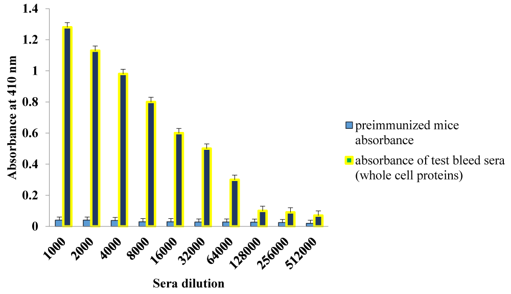
To evaluate the antibody titres raised in mice against Secretory proteins, total antibodies were measured. Sera samples were collected after third boosters. The cut-off value for the assays was calculated as the mean OD (+2 SD) from sera of control group assayed at 1∶1000 dilution. The endpoint antibody titers were proposed as reciprocal of the uppermost serum dilution giving an OD more than the cut-off. The antibody endpoint titer of Secretory proteins was 1.28×10⁵ which is shown in figure 2.
Figure 2: Depicted the Antibody titres against secretory proteins of C. botulinum type B (SP11) grown in CMM medium. Mice (n = 6 mice per group) were immunized with Secretory proteins or PBS as the control group. Four weeks after final immunization, antibody titre was measured with indirect ELISA. Results showed that titre against immunization of mice with Secretory proteins significantly increased compared with control group. Testing was carried out triplicate and values are shown as the mean ± S.D.
Detection limit
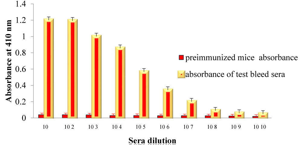
To develop an immunodetection system against C. botulinum type B, an indirect ELISA assay was used. For this assay, the antibodies were generated against secretory proteins of C. boulinum type B via intraperitoneal route. Polyclonal antibody against Secretory proteins was capable to detect C. botulinum type B approximately 106 cfu/ml. Detection limit is shown in figure 3.
Figure 3: Detection of C. botulinum type B with indirect ELISA using polyclonal antibody raised in mice against Secretory proteins. The error bars indicates the standard deviation of triplicates values.
Cytokine production levels
Cytokine profiles of secretory proteins anti -sera were determined by estimating the levels of GMCSF, IL-10 and IFN-γ. Significantly high expression levels of GMCSF and IFN-γ were noticed in antisera in comparison to PBS immunized mice serum. No significant difference was noticed in the expression levels of IL-10.
The most sensitive method available for the measurement of biologically active toxin is the mouse bioassay. The mouse bioassay is still considered the standard method for toxin detection and serotyping. To avoid animal use, a search for alternative in vitro assays of similar sensitivity is necessary. Immunoassays for botulinum neurotoxin detection are able of detecting as little as 10 to 100 minimum lethal doses/ml for type A toxin [11,12]. These in vitro methods were planned to virtually replace the mouse bioassay but have not been sufficiently validated for screening large numbers of samples. Ferreira et al. reported the use of an amplified ELISA for detection of preformed BoNT/A and culture toxins from hash brown potatoes associated with food-borne botulism [13]. But all above mentioned strategy are used for the detection of botulinum neurotoxins. But till now there is no detection system available for the detection of C. botulinum. In the present study, we developed the indirect ELISA to detect the C. botulinum type B. Secretory proteins were extracted from the C. botulinum type B. Antibodies were generated against the Secretory proteins in mice. Allergic diseases have been linked to Th2 immune responses, which are characterized by high levels of interleukins. These cytokines organize the enrolment and activation of dissimilar effector cells, such mast cells. These cells along with Th2 cytokines are main players on the development of chronic inflammatory disorders, typically considered by hyper responsiveness and airway inflammation. Accumulating indications have shown that changing cytokine-producing profile of Th2 cells by encouraging Th1 responses may be defensive against Th2-related diseases such as allergy and asthma. Interferon- γ the main Th1 effector cytokine, has revealed to be central for the resolution of allergic-associated immunopathologies. In the present study IFN-γ was expressed in high level [14]. GM-CSF act as no redundant function in the beginning of autoimmune inflammation irrespective of helper T cell polarization [15]. For indirect ELISA, C. botulinum type B was taken in different dilutions (cfu/ml). Generated polyclonal antibody was detected C. botulinum type B 106 cfu/ml. These findings are in agreement with other ELISA systems, where optimum reactions at a cell concentration of 106/ml or thereabouts has been achieved [16,17]. Merino et al were capable to detect as low as 10 cells per 100 ml of peptone water, which might have been due to the exact detector antibody against used by them [18]. Since there is no indigenous detection system available in the country, there is a need to develop an in-house system to detect botulism in clinical samples. So present study may be useful to detection of C. botulinum from clinical samples.
The author is thankful to the Director DRDE, Gwalior for providing necessary facilities and support throughout the work. I am also grateful to CSIR-UGC, India for their financial and worthy reward.
- Agarwal AK, Goel A, Kohli A, Rohtagi A, Kumar R. Food-borne botulism. J Assoc Physicians India. 2004; 52: 677-678. PubMed: https://www.ncbi.nlm.nih.gov/pubmed/15847370
- Chaudhry R, Dhawan VB, Kumar D, Bhatia R, Gandhi JC, et al. Outbreak of suspected Clostridium butyricum botulism in India. Emerg Infect Dis. 1998; 4: 506-507. PubMed: https://www.ncbi.nlm.nih.gov/pmc/articles/PMC2640317/
- Fillo S, Giordani F, Anselmo A, Fortunato A, Palozzi AM, et al. Draft genome sequence of Clostridium botulinum B2 450 strain from wound botulism in a drug user in Italy. Genome Announc. 2015; 3: e00238-15. PubMed: https://www.ncbi.nlm.nih.gov/pubmed/25838491
- de Jong LI, Fernández RA, Pareja V, Giaroli G, Guidarelli SR, et al. First report of an infant botulism case due to Clostridium botulinum type Af, J Clin Microbiol.2015; 53: 740-742. PubMed: https://www.ncbi.nlm.nih.gov/pubmed/25502535
- MacDonald E, Arnesen TM, Brantsaeter AB, Gerlyng P, Grepp M, et al. Outbreak of wound botulism in people who inject drugs, Norway, October to November 2013, Euro Surveill. 2013; 18: 20630. PubMed: https://www.ncbi.nlm.nih.gov/pubmed/24229788
- Hernández-de Mezerville M, Rojas-Solano M, Gutierrez-Mata A, Hernández-Con L, Ulloa-Gutierrez R et al. Infant botulism in Costa Rica: first report from Central America. J Infect Dev Ctries. 2014; 8: 123-125. PubMed: https://www.ncbi.nlm.nih.gov/pubmed/24423723
- Kautter DA, Solomon HM. Collaborative study of a method for the detection of Clostridium botulinum and its toxins in foods. J Assoc Off Anal Chem. 1977; 60: 541-545. PubMed: https://www.ncbi.nlm.nih.gov/pubmed/323214
- Cai S, Singh BR, Sharma S. Botulism diagnostics: from clinical symptoms to in vitro assays. Crit Rev Microbiol. 2007; 33: 109-125. PubMed: https://www.ncbi.nlm.nih.gov/pubmed/17558660
- Lindström M, Keto R, Markkula A, Nevas M, Hielm S, et al, Multiplex PCR assay for detection and identification of Clostridium botulinum types A, B, E, and F in food and fecal material. Appl Environ Microbiol. 2001; 67: 5694-5699. PubMed: https://www.ncbi.nlm.nih.gov/pubmed/11722924
- Sharma A, Ponmariappan S, Sarita R, Alam SI, Kamboj DV, et al. Identification of Cross Reactive Antigens of C. botulinum Types A, B, E & F by Immunoproteomic Approach. Curr Microbiol. 2018; 75: 531-540. PubMed: https://www.ncbi.nlm.nih.gov/pubmed/29332140
- Doellgast GJ, Beard GA, Bottoms JD, Cheng T, Roh BH, et al. Enzyme-linked immunosorbent assay-enzyme-linked coagulation assay for detection of antibodies to Clostridium botulinum neurotoxins A, B, and E and solution-phase complexes. J Clin Microbiol. 1994; 32: 105-111. PubMed: https://www.ncbi.nlm.nih.gov/pubmed/8126163
- Szílagyi M, Rivera VR, Neal D, Merrill GA, Poli MA. Development of sensitive colorimetric capture elisas for Clostridium botulinum neurotoxin serotypes A and B. Toxicon. 2000; 38: 381-389. PubMed: https://www.ncbi.nlm.nih.gov/pubmed/10669027
- Ferreira JL, Eliasberg SJ, Harrison MA, Edmonds P. Edmonds, Detection of preformed type A botulinal toxin in hash brown potatoes by using the mouse bioasssay and a modified ELISA test. J AOAC Int. 2001; 84: 1460-1464. PubMed: https://www.ncbi.nlm.nih.gov/pubmed/11601465
- Teixeira LK, Fonseca BP, Barboza BA, Viola JP. The role of interferon-gamma on immune and allergic responses, Mem Inst Oswaldo Cruz. 2005; 100: 137-144. PubMed: https://www.ncbi.nlm.nih.gov/pubmed/15962113
- Codarri L, Gyülvészi G, Tosevski V, Hesske L, Fontana A, et al. ROR [gamma] t drives production of the cytokine GM-CSF in helper T cells, which is essential for the effector phase of autoimmune neuroinflammation. Nat Immunol. 2011; 12: 560-567. PubMed: https://www.ncbi.nlm.nih.gov/pubmed/21516112
- Sachan M, Agarwal RK, A simple enzyme-linked immunosorbent assay for the detection of Aeromonas spp. Veterinarski Arhiv. 2002; 72: 327-334.
- Lee HA, Wyatt GM, Bramham S, Morgan MR. Enzyme-linked immunosorbent assay for Salmonella typhimurium in food: feasibility of 1-day Salmonella detection. Appl Environ Microbiol. 1990; 56: 1541-1546. PubMed: https://www.ncbi.nlm.nih.gov/pubmed/2200337
- Merino S, Camprubí S, Tomás JM. Detection of Aeromonas hydrophila in food with an enzyme‐linked immunosorbent assay. J Appl Bacteriol. 1993; 74: 149-154. PubMed: https://www.ncbi.nlm.nih.gov/pubmed/8444644


